Staphylococcus pseudintermedius ST2660 isolated from a cat has strong biofilm-forming ability and increases biofilm formation at cat’s normal body temperature
All experiments were performed in accordance with the relevant guidelines and regulations of the Biosafety Committee of Teikyo University School of Medicine.
Bacterial strains and growth conditions
Staphylococcus pseudintermedius (strain 2306K1) was isolated in 2023 from 8-year-old female cat with a severe wound abscess on the skin around the subcutaneous port of the SUB system. The cat was kept indoors with another cat and did not live with dogs. There was no history of antimicrobial treatment or infection in the past year before treating the cat. Bacteria in a specimen was identified by Sanritsu-Zelkova Inspection Center (Tokyo, Japan) and the isolate was reidentified by MALDI-TOF MS using the MALDI Quick Microbiological Identification Test (ICLAS Monitoring Centre, Central Institute for Experimental Medicine and Life Science, Kanagawa, Japan). S. aureus (strain ATCC 25923) and S. haemolyticus (strain 1369 A) were used as the control strain biofilm in this study36,37. These strains were stored in glycerol at -80 °C in the Department of Microbiology and Immunology, Teikyo University School of Medicine. These bacteria were cultured on LB agar plates (Becton, Dickinson and Company, Franklin Lakes, NJ, USA) for 18 h at 37 °C. Thereafter, the bacteria were suspended in LB broth and the cell concentration was determined by measuring the optical density (OD) at 595 nm. The resulting bacterial suspensions were used for subsequent experiments.
MLST analysis
DNA was extracted from the S. pseudintermedius strain 2306K1. Seven loci (ack, cpn60, fdh, pta, purA, sar, and tuf) were amplified using PCR and sequenced using a DNA sequencing service (Eurofins Genomics, Tokyo, Japan). The primer sequences are listed in Supplementary Table S1. The PCR was performed as follows: 35 denaturation cycles at 95 °C for 15 s, annealing at 60 °C for 30 s (cpn, pta, fdh, ack, and sar), annealing at 56 °C for 30 s (tuf and purA), and extension at 72 °C for 1 min. ST was assigned using MLST, as assessed by the Oxford scheme, in the PubMLST database.
PCR screening for virulence factors in S. pseudintermedius strain 2306K1
To determine the presence of genes encoding toxins and two pivotal genes from ica locus related to biofilm formation, the target genes (lukS and lukF for cytotoxins; siet for exfoliative toxin; sel, sem, and seq for enterotoxins; icaA and icaD for biofilm regulation) were screened using PCR reactions. The primer sequences are listed in Supplementary Table S1. The PCR was performed as follows: 35 denaturation cycles at 95 °C for 15 s, annealing at 55 °C for 30 s, and extension at 72 °C for 1 min.
Determination of MIC
Antimicrobial susceptibility testing was performed according to the CLSI guidelines, CLSI M100-ED34:2024 or CLSI VET01S ED7:2024. Antimicrobial susceptibility testing was performed using a KB disk (Eiken Chemical Co. Ltd., Tokyo, Japan), and the MIC was determined using the broth microdilution method in CAMHB (Becton, Dickinson and Company) and LB broth (Sigma-Aldrich, St. Louis, MO, USA). Amoxicillin, clavulanic acid, and doxycycline were purchased from Merck KGaA (Darmstadt, Germany), and cefalexin was from FUJIFILM Wako Pure Chemical Corporation (Osaka, Japan).
Determination of biofilm-forming ability
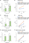
The growth of planktonic, biofilm-dispersed, and biofilm cells of S. pseudintermedius strain 2306K1, S. aureus strain ATCC 25923, and S. haemolyticus strain 1369A was analysed using a biofilm formation assay kit (DOJINDO LABORATORIES, Kumamoto, Japan)38. These bacteria, adjusted to 4 × 10⁵ CFU/mL in LB broth, were added to a 96-well microtitre plate provided by the kit. Then, the plate was covered with a 96-peg microtiter plate lid (96-peg lid) and incubated for 24 and 48 h at 37 and 39 °C in each combination. These bacteria were cultured at 39 °C because the average normal cat body temperature is approximately 37.7–39.5 °C39. After incubation, the 96-peg lid was removed, and the culture broth was determined by measuring OD595 to analyse the growth of planktonic and biofilm-dispersed bacterial cells. To analyse the biofilm cells of these bacteria, the 96-peg lid was washed two times with sterile PBS and placed onto a fresh 96-well microtitre plate, with each well containing 0.1% crystal violet solution. After staining for 30 min, the 96-peg lid with the stained biofilm cells was washed two times with sterile PBS to remove unreacted crystal violet. The 96-peg lid was then placed onto a fresh 96-well microtitre plate, with each well containing 99.5% ethanol, and extracted for 15 min. The quantity of extracted crystal violet was determined by measuring the absorbance at 590 nm (Abs590). The BFI was calculated as [BFI = (Abs590 (biofilm cells) / OD595 (planktonic or biofilm-dispersed cells)], providing the extent of biofilm formation.
Determination of inhibitory and bactericidal effects of amoxicillin/clavulanic acid, cefalexin, and doxycycline on planktonic, biofilm-dispersed, and biofilm cells of S. pseudintermedius strain 2306K1
The inhibitory and bactericidal effects of amoxicillin/clavulanic acid, cefalexin, and doxycycline on planktonic, biofilm-dispersed, and biofilm cells of S. pseudintermedius strain 2306K1 were analysed using biofilm formation and viability assay kits (DOJINDO LABORATORIES)38. The strain 2306K1, adjusted to 4 × 10⁵ CFU/mL in LB broth using various concentrations of these antibiotics, was added to a 96-well microtitre plate provided in the kit. Then, the plate was covered with a 96-peg microtitre plate lid (96-peg lid) and incubated for 24 h at 37 and 39 °C. After incubation, the 96-peg lid was removed and the culture broth was measured at OD595 to analyse the bacterial growth of planktonic cells and determine the inhibitory effects of these antibiotics. To analyse the bactericidal effects of these antibiotics on the planktonic cells, 5 µL of culture broth was plated on LB agar plates, and the presence of viable cells was confirmed through the growth of the bacterial suspension after 24 h of incubation at 37 °C. The MBC was defined as the lowest concentration at which the bacteria were killed after 24 h of incubation. To analyse the inhibitory effects of these antibiotics on the biofilm cells of strain 2306K1, the 96-peg lid was washed two times with sterile PBS and then placed onto a fresh 96-well microtitre plate, with each well containing 0.1% crystal violet solution. After staining for 30 min, the 96-peg lid with the stained biofilm cells was washed two times with sterile PBS to remove unreacted crystal violet. The 96-peg lid was then placed onto a fresh 96-well microtitre plate, with each well containing 99.5% ethanol, and extracted for 15 min. The quantity of crystal violet extracted was determined by measuring Abs590. The viability of the biofilm cells of strain 2306K1 was determined using a biofilm viability assay kit according to the manufacturer’s protocol. Briefly, after bacteria were cultured in a 96-well microtitre plate covered with a peg lid, the 96-peg lid was washed two times with sterile PBS to remove planktonic and non-attached cells and then placed onto a fresh 96-well microtitre plate with each well containing LB broth and 2-(4-Iodophenyl)-3-(4-nitrophenyl)-5-(2,4-disulfophenyl)-2 H-tetrazolium monosodium salt (WST-1) solution in a biofilm viability assay kit. After incubation for 24 h, the 96-peg lid was removed and the absorbance of the LB broth was measured at Abs450.
To analyse the inhibitory and bactericidal effects of amoxicillin/clavulanic acid, cefalexin, and doxycycline on the biofilm-dispersed and biofilm cells of strain 2306K1, the bacteria were added to a 96-well microtitre plate provided in the kit. The plate was covered with a 96-peg microtitre plate lid and incubated for 24 h at 37 and 39 °C. After incubation, the 96-peg lid with the established biofilm cells was washed two times with sterile PBS and placed onto a fresh 96-well microtitre plate, with each well containing LB with various concentrations of antibiotics. Subsequently, after incubation for 24 h at 37 and 39 °C, the inhibitory and bactericidal effects of antibiotics on the biofilm-dispersed and biofilm cells of strain 2306K1 were analysed using the previously presented methods. The MIC, MBIC, and MBEC were defined as > 95% inhibition or eradication relative to the untreated sample.
Scanning electron microscope
Biofilms grown on the slide glass fragment (SPL Life Sciences, Korea) in a 96-well plate were rinsed with PBS (three times, 10 min each) and subsequently fixed in mixed solution including both 2% glutaraldehyde solution (FUJIFILM Wako) and 2% formaldehyde (Becton, Dickinson and Company) at 37 °C for 15 min, then at 25 °C for 10 min, and finally at 4 °C for 5 min. After the cells were rinsed with double distilled water (three times, 10 min each), the cells were treated with 1% tannic acid (FUJIFILM Wako) at 4 °C for 15 min. Following fixation, biofilms on the slide glass fragment were rinsed with double distilled water (three times, 10 min each), post-fixed in 2% osmium tetroxide (FUJIFILM Wako) at 25 °C for 30 min, rinsed in double distilled water (three times, 10 min each), and dehydrated in a graded series of ethanol (50% [10 min], 70% [10 min], 80% [10 min], 90% [10 min], 95% [10 min], and 100% [twice, 5 min each; once, 15 min; once, 30 min]). After dehydration in ethanol, the biofilms were substituted with a graded series of t-butyl alcohol (50% [10 min], 75% [10 min], and 100% [twice, 5 min each; once, 20 min]). The samples were freeze-dried (vacuum device, VFD-21 S, Ibaraki, Japan), sputter-coated with platinum (Pt) (vacuum device, MSP-20-UM), and imaged using a scanning electron microscope (Hitachi TM-3030, Hitachi High-Tech Corporation, Tokyo, Japan) operating at 15.0 kV in the charge reduction mode.
Statistical analysis
Results for the growth of planktonic, biofilm-dispersed and biofilm cells of the bacteria were compiled from two independent experiments and are presented as the mean ± standard error of the mean (SEM). Results for the planktonic, biofilm-dispersed, and biofilm cells of the bacteria in the presence of antibiotics were combined from two independent experiments and presented as the mean ± SEM. Numerical data were compared using one- and two-way factorial analyses of variance (ANOVA), followed by Bonferroni’s multiple comparison test. A two-tailed P value set than 0.05 was considered significant.


